Wednesday, February 22, 2012

Plantar Fasciitis - The Leading Cause of Heel Pain

A common condition that affects the foot is heel pain. The characteristic pain affecting the heel is variable. It can be described as burning pain, slow progressing pain, or striking pain when the affected heel is used. In some cases, especially when a patient has chronic heel pain, the pain can occur even at rest.
The plantar fascia is involved in most cases of heel pain. There could be a pathologic finding or damage to plantar fascia or irritation and inflammation to the surrounding tissues at the heel bone where the plantar fascia is attached.
The plantar fascia is a powerful band of fibrous tissue found underneath the foot. It joins to the calcaneus or heel bone to the balls of the foot. It is involved in maintaining the arch of the foot which is essential to the biomechanics of the foot.
The plantar fascia is constantly under extreme pressure when walking, running, lifting weights, or anything that involves using the foot. Because of this, it is not surprising that this structure develop tiny tears that can accumulate over the years. These tiny micro tears can cause mild to severe heel pain. This is the reason why older adults commonly developt this type of foot pain. Younger individuals can also develop premature fascia tears especially those who are active in sports like runners. Plantar fasciitis is the medical term which means irritation and inflammation of the plantar fascia.
Heel pain is a very common condition. One out of ten people will experience at least one episode of heel pain at some point in their lives. People mostly affected by heel pain are:
  • The elderly around the age 40 to 60 years old.
  • Active individuals who regularly run or jog.
  • Athletes like runners, basketball players, volleyball players.
  • Obese or overweight
  • Diabetics
Treatment of heel pain due to plantar fasciitis focuses on alleviating the inflammation and irritation of the plantar fascia. The simplest way to manage heel pain is by following the R.I.C.E.. method.
  • Immobilizing and resting the foot promotes wound healing. Resting alone can manage the acute or early stages of heel pain. Resting alone can treat the acute or early stages of heel pain. This heals the micro tears and damage done to the plantar fascia
  • Ice. Cold stimuli help reduce inflammation and can also decrease pain
  • Compression. Compression bandages help immobilize the foot and also reduce further swelling of the foot
  • Elevating the foot above the heart prevents blood pooling. This significantly reduce swelling therefore also an effective method to decrease pain. This significantly reduce swelling therefore also a great way to reduce pain
Other treatments include calf stretches, steroid injections, devices that support the foot like orthoses or shoes that provide cushion and support for the foot.
If the above treatments still do not alleviate pain, then other modalities like ESWT or minimal surgery is required especially for chronic heel pain.

Monday, February 20, 2012

Fungus Among'st Us

Nail fungus is a slow moving colonization of human keratin, which rarely causes life threatening issues, but may almost always cause some form of discomfort with shoe gear, ambulatory compromise, or unsightly appearance to the feet.

Nail fungus attacks all people. But there are some people that have higher risk of getting nail fungus infection. Who are those people, and why the more risk?

In neral, this applies with people that have lower immune system. This includes people with circulatory system problem, diabetic people, late-aged people and patients with cancer and chemotherapy treatment, for instance. Also with age, comes the likelihood of increased fungal risk. Low immune systems may allow the fungus to spread easily. Usually, once it infects one toenail or nail, they will spread immediately to the other nails on the same feet or hand over many years.

There are many kinds of treatments; ranging from mild to aggressive. Some used home-made remedies, or some use natural treatments, and some visit the doctor. The latter option is the one that most people choose, because they want effective and safe treatment. But what they don't know is that prescribed medicines can cause side-effects because of their toxic chemical ingredients. This can be hard for people with liver diseases, because nail fungus treatment can take 3 months or more.

Foot care centers use a variety of topical therapy, and have had excellent success with the Cool Touch Nail Laser System. The success rate statistics of treatment is 70 to 80 percent. There is a 15% chance that it will reoccur. Because of this statistic, many doctors advise the use of anti-fungal solutions after treatment to prevent another infection.

Simple tips to prevent fungus infection:

Wear open footwear as much as possible.

Change socks immediately if it becomes damp or wearing absorbent socks is more preferable.

Don't wear high top boots if not needed.

Treat Nail Fungus as early as you can to stop its spreading.

Family Foot and Leg Center
is a medical center specializing in healing health issues with lower leg and ankle. Come to us to get treatment for all your leg disorders or obtain information about our practices.

Common Causes of Heel Pain

When walking, our feet are always under stress. The truth is, our feet can carry heavy loads but excessive stress can push them to their limits. When you're involved in sports like running, playing basketball or volleyball and even using the wrong shoes can irritate tissues in the foot that can lead to heel pain.

When there is tissue inflammation around the heel, this will cause considerable amount pain on the affected area. This can heal itself without any medication if the foot is immobilized and allowed to rest. Sadly, a lot of people ignore the early signs of heel pain and go on with their usual activities. This can ultimately lead to chronic heel pain and will need one or more treatment modalities to alleviate pain.

Diagnosing heel pain and it's causes are best done by a podiatrist. Recognizing the cause is crucial because there may be different or similar treatment options for each condition.

Mainly, there are two categories in the diagnosis of heel pain. These are posterior heel pain or pain at the back of the heel and inferior heel pain or pain underneath the foot.

Posterior heel pain usual involves the insertion of the Achilles tendon to the calcaneus or heel bone. This is common among runners or in people who wear shoes that can apply pressure at the back of the foot. This initially starts are tolerable pain and can get worse if ignored.

Inferior heel pain involves an irritated plantar fascia. The plantar fascia is a strong fibrous tissue found underneath the foot. Same as the Achilles tendon, the plantar fascia also inserts to the heel bone to the digits of the feet. The plantar fascia is constantly under pressure when walking, running, or lifting weight. It also plays a vital role in maintaining the arch of the foot. Plantar fasciitis is the term used for an inflamed or irritated plantar fascia. Irritation occurs at the insertion of the plantar fascia to the heel bone. In chronic plantar fasciitis, it is common to find a heel spur on radiologic findings.

Treatment involves the R.I.C.E method, which stands for rest, ice, compress, and elevate. This is very effective in the early course of heel pain. On the other hand, chronic heel pain is more difficult to manage especially when a spur has already formed. It is best to visit a podiatrist when dealing with chronic pain.

For more information about heel pain, please visit the Family Foot and Leg Center in Naples or Contact Dr. Kevin Lam at (239) 430-36

Monday, February 6, 2012

Understanding the Development of A Heel Spur

What is a heel spur?

A heel spur is a bony formation located at the calcaneous bone or heel bone that is evident through x-ray. Also called calcaneal spur, this condition is commonly associated with plantar fasciitis when the spur is located at the inferior part or at the bottom of the calcaneous bone.

Though these conditions are related, they are two separate disease entities. Plantar fasciitis refers to the irritation or inflammation of the plantar fascia, and this is sometimes accompanied by a spur. But not all patients with plantar fasciitis develop a heel spur and not all patients with heel spurs have plantar fasciitis.

Heel spurs are seen on patients who have chronic heel pain due to the inflammation of the plantar fascia. This is often seen on middle aged men and women or among active athletic individuals involved in sports like runners, basketball players and volleyball players.

Patient don't actually complain of heel spur pain but pain from plantar fasciitis. Most often, a heel spur can only be found or diagnosed through radiological findings where the pointy bony prominence is located at the attachment of the plantar fascia.

How do heel spurs form? About 70% of patients with plantar fasciitis will develop heel spurs. The plantar fascia, which is located at the bottom of the foot, is a strong fibrous tissue that attaches to the heel bone to the balls of the foot. It is responsible for the arch of the foot and plays a major role in the biomechanics of the foot. This fascia is also under enormous stress when running, walking or bearing weight.

However, this important structure is very prone to injury and degenerative changes. The natural wear and tear of this tendon can cause irritations and inflammation causing chronic heel pain. For some reason, the new bone growth is formed at the attachment of the plantar fascia to the heel bone probably because of the constant stressful pulling action.

Treating pain associated with heel spurs and plantar fasciitis can be done conservatively. Most often, the R.I.C.E. method which stands for rest, ice, compress, and elevate can effectively relive pain. Other treatments are foot inserts, pain medications and steroid injections.

A new treatment called ESWT uses multiple shock waves to naturally stimulate healing in the injured fascia. This is a holistic approach since it encourages the bodies own healing properties.

For more information about heel spurs and plantar fasciitis, please visit the Family Foot and Leg Center in Naples or Contact Dr. Kevin Lam at (239) 430-3668.

Tuesday, January 24, 2012

Information About Gout - Signs and Symptoms


Gout is a very common type of arthritis characterized by sudden attacks of burning pain, reddening, swelling and warm/heat felt from the affected area. About 50% of gout cases affect the big toe, though it can also affects other joints like the ankle, this areas are a major concern since ankle or heel pain can be incapacitate and can affect the well being or a patients. Gout is a chronic condition and pain can anytime without prior warnings or signs. If not properly treated, this can damage joints, tendons and the surrounding tissues.

The cause of gout is due increased amounts of circulating uric acid in the blood. This blood uric acid overload or hyperurecimia, produces sharp pointy urate crystals that can deposit in the joints and can form into large lumps causing severe pain. The urate crystals can form in other organs as well especially in the kidneys and can lead to kidney stone formation or renal failure due to urate deposits in filtering tubules.

There are about 5 million Americans are affected by gout. Men are 9 times more prone to gout than women. Typically, gout in men starts in puberly. In women, it usually starts after menopause.

The risk factors involving gout formation are:

Obesity
Increase alcohol intake
Hypertension
Renal or kidney dysfunction
Increased intake of purine rich foods (anchovies, canned good, processed meats)
Certain medications: diuretics, anti-TB drugs, aspirin

The signs and symptoms of gouty arthritis involve the four classic signs of inflammation:

Color warmness or heat felt in the inflamed area
Dolor- pain and tenderness
Rubor redness
Tumor swelling

Fever is also a symptom in gouty arthritis that usually occurs on acute attacks.

High suspicion of gouty arthritis involves a history of joint pains especially in the toes. Oddly, gout pain always involve one joint at a time, as compared to multiple joint pains in other arthritic conditions. Finding urate crystals on joint fluids by needle aspiration is the mainstay for diagnosing gout.

Family Foot and Leg Center in Naples, FL offers holistic approaches on treating gouty arthritis involving the big toe or ankle. Contact Dr. Kevin Lam at (239) 430-3668 for more details.

GouTrol is a physician formulated supplement to help with the symptoms of gout.

Sunday, January 15, 2012

Do You Have a Hammer Toe?


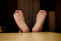
When the smaller toes of the foot become bent and prominent these are called hammer toes. The 4 smaller toes of the foot are very much like the same fingers in the hand. Each has three bones (phalanges) which have joints between them (interphalangeal joints). The toes form a joint with the long bones of the foot (metatarsals) and it is this area that is often referred to as the ball of the foot.

Generally, these bones and joints are straight. A hammertoe occurs when the toes become bent at the first interphalangeal joint, making the toe prominent. This can affect any number of the lesser toes. In some cases, a bursa (rather like a deep blister) is formed over the joint and this can become inflamed (bursitis). With time, hard skin (callous) or corns (condensed areas of callous) can form over the joints or at the tip of the toe.

What causes hammertoes?

There are many different causes but commonly it is due to shoes or the way in which the foot works (functions) during walking. If the foot is too mobile and / or the tendons that control toe movement are over active, this causes increased pull on the toes which may result in deformity.

In some instances trauma (either direct injury or overuse from walking or sport) can predispose to hammertoes. Patients who have other conditions such as diabetes, rheumatoid arthritis and neuromuscular conditions are more likely to develop hammertoes.

Are women more likely to get the problem?

It is more common in women because they tend to wear tighter, narrower shoes with increased heel height. These shoes place a lot of pressure onto the joint and predispose to deformity. It is common for patients to wear shoes that are too small and this can predispose to the problem. In a study we made, about 95% of patients were in the wrong size shoes.

Will it get worse?

At the start of the deformity, it is generally mobile which means that the toe can be straightened. However, with time, the joint become rigid or fixed. This can then affect the joint at the ball of the foot and, in severe cases, the joint capsule ruptures (tears) so that the joint becomes dislocated and the toe sits up in the air.

What are the common symptoms?

  • Deformity / prominence of toe
  • Pain
  • Redness around the joints
  • Swelling around the joints
  • Corn / Callous
  • Difficulty in shoes with deformity of the shoe upper
  • Difficulty in walking
  • Stiffness in the joints of the toe

How is it identified?

Clinical examination and a detailed history allow diagnosis. X-rays are often not required but can help to evaluate the extent of the deformity and the degree of arthritis within the joint.

What can I do to reduce the pain?

There are several methods that you can do to relieve your symptoms:

  • Wear good fitting shoes with a deep toe box
  • Avoid high heels
  • Use a toe prop to straighten the toe if it is still mobile
  • Wear a protective pad over the toe
  • See a doctor at the Family Foot and Leg Center

As a specialist, what we can do to correct or reduce your symptoms?

If simple measures do not reduce your symptoms, there are other options:

  • Advise appropriate shoes
  • Advise exercises if the toes are still mobile
  • Show you how to strap the toe in a corrected position
  • Provide a splint or protection
  • Consider orthotics

Advise on surgery

The way in which your foot loads during walking can place increased stress on the ball of the foot and cause increased toe activity. Special shoe inserts (orthoses) can help to control foot movement. Whilst these are unlikely to resolve established deformity they may help reduce discomfort in the ball of the foot.

Will this cure the problem?

If the deformity is mobile, then this may help prevent progression although there have been no scientific studies to analyse the benefit. If the deformity is fixed, then orthotics will not cure the problem but may reduce the associated symptoms.

What will happen if I leave this alone?

Generally, the deformity becomes worse with time and slowly becomes fixed (stiff). This can cause discomfort in shoes. The position of the toe places increased stress on the ball of the foot and this can become painful. Corn and callous formation on the ball of the foot is not uncommon. In some cases, the metatarsophalangeal joint capsule ruptures, causing the toe to sit up in the air.

Can the deformity be reversed or cured?

The only effective way of correcting the deformity is to have an operation.

How does the operation correct the deformity?

There are a number of different operations. However, the most common operations are:

  • Tendon transfer
  • Digital arthroplasty
  • Digital arthrodesis

Tendon transfers involve taking the tendon from under your toe and re-routing it to the top of the toe so that the toe is pulled down. This can be used alone if the toe is mobile or in combination with the other two procedures. This can leave the toe a bit swollen and stiff.

Digital arthroplasty and arthrodesis involve the removal of bone from the bent joint to allow correction. An arthroplasty removes half the joint and leaves some mobility whilst an arthrodesis removes the whole joint and, following a period of time with a wire/pin protruding from the end of the toe, leaves the toe rigid.

In more severe cases, the tendon on the top of the toe and the joint at the ball of the foot need to be released to allow the toe to straighten. If there is severe stiffness at this joint, then the base of the bone at the bottom of the toe (phalanx) may need removing (basal phalangectomy) or the metatarsal shortened (Weil osteotomy).

Patients will often tell me this: "I have heard it is very painful."

The nature of surgery means that there will be pain and swelling, usually worse the night after surgery. However, with modern anaesthetic techniques and pain killers, this can be well controlled. The level of pain experienced varies greatly from patient to patient with some experiencing no significant discomfort.

Will I have to have a general anaesthetic (be asleep)?

Not if you did not want one. All of these procedures are done perfectly safe while your awake, under local anestetic. Some patients worry that they may feel pain during the operation but it would not be possible to perform the operation if this were the case. We often perform these procedures at our surgical suite over at the Gridley Building location, where often times these procedures are done within 30 minutes, and you leave right then in a surgical shoe with the dressing applied immediately after the procedure is completed.

Will I have to stay in hospital?

No. As long as you were medically fit and have adequate home support, many patients are able to have this type of operation performed as day surgery and go home.

Will I have to have a plaster cast?

Plaster casts are generally not required for this type of surgery.

Are there a lot of complications?

All operations have complications and risks and these should be discussed in detail with your specialist. However, with most foot surgery it is important to remember that you may be left with some pain and stiffness and the deformity may reoccur in the future. This is why it is not advisable to have surgery if the deformity is not painful and does not limit your walking. A thorough examination of your foot and general health is important so that these complications can be minimised.

Although every effort is made to reduce complications, these can occur. In addition to the general complications that can occur with foot surgery, there are some specific risks with toe surgery:

  • Persistent swelling which may be permanent
  • Recurrence of deformity / corn (this tends to be more of a problem with the little toe)
  • Regrowth of removed bone
  • Residual pain
  • Stiffness or flail (floppy) toe
  • The toe may not sit on the ground floating toe (there is an increased risk of this with arthrodesis)
  • You may get discomfort in other parts of your foot during the recovery period. This generally settles.
  • There is always a possibility that the deformity may return in later life.

When will I be able to walk again and wear shoes?

In the majority of cases, you will able to walk with the aid of crutches within 2-4 days but you will remain somewhat limited for the first 2 weeks.

Some patients are able to return to wider shoes within two weeks with 60% of patients in shoes at 6 weeks and 90% in 8 weeks. This period is longer for arthrodesis as shoes cannot be worn until the wire/pin has been removed (generally 3-6 weeks).

Swelling generally starts to reduce at 6-8 weeks and the foot will be beginning to feel more normal at 3 months although the healing process continues for 1 year.

When will I be able to drive again?

When you feel able to perform an emergency stop. This is generally between 4-8 weeks post operatively but you should always check with your insurance company first.


When will I be able to return to work?

If you are able to get a lift and have a job that is not active and you can elevate your foot, you may be able to return after 1-2 weeks. Generally, patients return to work between 4-8 weeks depending on the type of job, activity levels and response to surgery.

When will I be able to return to sport?

Although the healing process continues for up to 1 year, you should be able to return to impact type activity at around 3 months. This will depend on the type of operation you have and how you respond to surgery.


Overview of Peroneal Tendon Problems

Overview of Peroneal Tendon Problems

Many patients experience chronic ankle instability in the form of lateral tendon dislcocations, or chronic ankle sprains. Many times these patients are unclear as to how important these tendons are to the overal stability and function of the ankle joint. With abnormal tendon gliding and ligamentous attenuations and ruptures, these tendons may also become painful with patients who have chronic ankle sprains. This is a comprehensive overview of this pathology and treatment options to help out with the understanding of these clinical scenarios.

History of the Procedure


Peroneal tendon abnormaliity have been reported uncommon. Monteggia described peroneal tendon subluxation in 1803, and this entity seems to be more commonly encountered than are disruptions of the peroneus longus or brevis alone. Nonetheless, peroneus brevis disorders have been described more often in the literature, with peroneus longus problems gaining more recent attention. Nonetheless, much of the literature regarding both tendons is in the form of case reports.

Problem


The peroneal muscles make up the lateral compartment of the leg and receive innervation from the superficial peroneal nerve. The peroneus longus muscle originates from the lateral condyle of the tibia and the head of the fibula. The tendon of peroneus longus courses behind the peroneus brevis tendon at the level of the ankle joint, travels inferior to the peroneal tubercle, and turns sharply in a medial direction at the cuboid bone. The tendon inserts into the lateral aspect of the plantar first metatarsal and medial cuneiform.

A sesamoid bone called the os peroneum may be present within the peroneus longus tendon at about the level of the calcaneocuboid joint. The frequency with which an os peroneum occurs is controversial, with many supporting the idea that one is always present. However, the os peroneum may be ossified in only 20% of the population. The peroneus longus serves to plantar flex the first ray, evert the foot, and plantar flex the ankle.

The peroneus brevis originates from the fibula in the middle third of the leg. Its tendon courses anterior to the peroneus longus tendon at the ankle. It courses over the peroneal tubercle and inserts onto the base of the fifth metatarsal. The peroneus brevis everts and plantar flexes the foot.

Problems may arise in either of the tendons alone, or both may be involved with subluxation. The hallmark of disorders of the peroneal tendons is laterally based foot or heel pain. Whether the problem is tendinous degeneration or subluxation, the clinical manifestation is pain. With time, loss of eversion strength may occur.

Problems arising with the peroneus longus include tenosynovitis and tendinous disruption (acute or chronic). The os peroneum may be involved with the degenerative process or as a singular disorder and can be fractured or fragmented. Longitudinal tears of the peroneus longus are uncommon but have been reported.

Longitudinal tears of the tendon are the most common problem seen with the peroneus brevis tendon. These may be single or multiple. Tendinitis and tenosynovitis also may occur.

Subluxation of both peroneal tendons may occur following an acute traumatic episode or may be of a more chronic nature.

Frequency


Disorders of the peroneal tendons are less common than other tendon problems involving the Achilles or posterior tibial tendons. However, it is impossible to estimate their true frequency in the United States or abroad.

Etiology


The precise etiology of peroneal tendon disorders depends somewhat on the specific problem being addressed. All disorders may result following a traumatic episode, direct or indirect, with a lateral ankle sprain being the most common trauma. Brandes and Smith have reported that 82% of patients with primary peroneus longus tendinopathy had a cavo-varus hindfoot.3 The presence of an os peroneum also has been postulated to predispose to peroneus longus rupture. Ruptures likewise have been reported to occur secondary to rheumatoid arthritis and psoriasis, as well as diabetic neuropathy, hyperparathyroidism, and local steroid injection.4,5,6

Longitudinal splits in the peroneus brevis tendon appear to result from mechanical factors. Repetitive or acute trauma causes the attritional ruptures. These ruptures may result from an incompetent superior peroneal retinaculum that allows the peroneus brevis to rub abnormally against the fibula.

Overcrowding from a peroneus quartus muscle also has been reported. The blood supply to the tendon has been shown to be adequate.

Subluxation of the peroneal tendons results from disruption of the superior peroneal retinaculum and usually involves avulsion of the retinaculum from its fibular insertion. The mechanism of injury typically involves an inversion injury to the dorsiflexed ankle with concomitant forceful contraction of the peroneals. Some patients have a more chronic presentation and cannot recall a traumatic episode. Congenital dislocations also have been reported. An inadequate groove for the peroneals in the posterolateral fibula may be a cause of subluxation as well.

Pathology of the longus and brevis tendons almost always occurs concurrently. Brandes and Smith noted a 33% incidence of concomitant problems.

Pathophysiology


Brandes and Smith have described and classified primary peroneus longus tendinopathy.3 They present 3 anatomic zones in which the tendon can be injured. Zone A is the level of the superior peroneal retinaculum. Zone B is the level of the inferior peroneal retinaculum. Zone C is the level of the cuboid notch. In their series, complete ruptures were most likely in zone C, while partial ruptures were more common in zone B. In the same study, surgical findings were classified into 3 groups. Group I pathology had no frank rupture but did have adhesion's or thickening of the tendon. Group II pathology consisted of partial tears with some continuity of the tendon. Group III had complete ruptures with complete loss of continuity. All group III pathology occurred in zone C.

Other attempts have been made to classify peroneal tendon pathology. Sobel et al have presented a classification for tears of the peroneus brevis tendon as follows:7,8

Grade 1 - Flattened tendon
Grade 2 - Partial-thickness split less than 1 cm in length
Grade 3 - Full-thickness split less than 2 cm in length
Grade 4 - Full-thickness split more than 2 cm in length

Eckert and Davis have classified superior peroneal retinaculum (SPR) pathology as follows:9

Grade I - SPR elevated from fibula
Grade II - Fibrocartilaginous ridge elevated from fibula with SPR
Grade III - Cortical fragment avulsed with SPR

Presentation

The patient with peroneal tendon pathology typically complains of laterally based ankle or hindfoot pain. The pain usually worsens with activity. However, presentation and diagnosis often are delayed. Patients may or may not recall a specific episode of trauma. Brandes and Smith reported that only 9 of 22 patients with primary peroneus longus tendinopathy recalled an inciting event and that the event was an average of 4.3 months prior to presentation.

Peroneal tendon subluxation or dislocation may present acutely following a traumatic injury to the ankle. However, it is not uncommon for these to present later with an uncertain history of trauma. Patients also may complain of snapping or popping in the ankle.

On physical examination, there usually is tenderness to palpation along the course of the peroneal tendons. Edema also may be present. These disorders require a high level of suspicion. Even frank dislocations may be missed if not specifically evaluated.

A provocative test for peroneal pathology has been described. The patient's foot is examined hanging in a relaxed position with the knee flexed 90ยบ. Slight pressure is applied to the peroneal tendons posterior to the fibula. The patient is then asked to forcibly dorsiflex and evert the foot. Pain may be elicited, or the tendons may be felt to sublux.

Indications

The primary indication for treating these disorders is pain. Nonsurgical treatment usually is attempted first. Failure of conservative measures is an indication for operative intervention.

Operative Considerations

With physical therapy, MRI, and need for primary or secondary repair will be determined based on overall health of the patient, as well as how effective nonsurgical measures have been. If the pain and resolution is not fully noted through physical therapy and bracing one should consider the possibility of repair. If there are tendon tears associated with the pathology, surgical repair is recommended. If an associated ruptured retinaculum or low muscle (peroneus quartius) is identified, surgical repair is also likely required. We are experts in this pathology, and treat this regularly, and I feel a proper evaluation for this condition will be beneficial to anyone with recurrent ankle sprains, as well as pain in the lateral ankle.

Visit us at naplesheelpain

Tuesday, January 10, 2012

Relieve Heel Pain with RICE

40+60 Feet, Euw.Image by bark via FlickrHeel pain a common condition that affects adults of all ages. The heel bone is the largest bone of the foot. For every step taken, the lower portion of the heel bone is the first part of the foot that hits the floor. Our foot is specially disigned for walking, but injuries can still happen if we don't practice proper foot care.

Plantar fasciitis is the most common cause of heel pain. This is due to the irritation or inflammation of the plantar fascia.

The plantar fascia is located under the foot. It is a strong fibrous ligament that is responsible for the arch of the foot and serves a very important role for the foot's biomechanics.

Injury of the plantar fascia is due to several factors like sports (especially in athletes involved in sprinting), using improper footwear, and being overweight to name a few. These injuries can cause the fascia to become inflamed creating variable intensity of pain in the foot.

Treatment of heel pain is easily managed at home. The best modality is to follow the R.I.C.E. method which stands for rest, ice, compress, and elevation.

1. Resting the foot is the initial step. Resting the affected area helps heal any injuries of the foot. Total immobilization is recommended for one to two days. It is also advised not to take any pain killers during this period because this masks the healing process since it "temporarily" alleviates pain, tempting you to use your foot.

2. Ice packs works great in releaving severe pain, it also help reduce swelling. Apply the ice packs every hour for 20 minutes.

3. Compression helps in controlling the swelling. Lightly wrap the elastic bandage around the affected area.

4. Elevating the foot helps prevent swelling and assists in venous return to the heart.

If heel pain still persists after the R.I.C.E. method, see your foot specialist.
Enhanced by Zemanta

All About Foot and Ankle Infections


This is a relatively common medical condition seen at the Family Foot and Leg Center in Naples, Florida. Many patients will end up with this devastating diagnosis and it will lead to a number of amputations across the country, with each year steadily increasing the number of foot and leg amputations primarily from patients with diabetes and the associated complications from such especially with neuropathy and vascular disease.

We try to utilize the most innovative technology to try and prevent this devastating complication (i.e. amputations) , with the use of state of the art external fixators, MicroVas microcirculatory therapy, wound care referrals, and closely working with infectious disease doctors for management of the systemic portion of treatment especially in the case of ankle osteomyelitis. This commonly includes intravenous antibiotics and weekly monitoring of blood tests. There are cases, the foot, feet or even leg are partially amputated.

FFLC prides themselves on diabetic limb salvage, which essentially includes treatment of bone infections of the ankle and foot, as well as prevention of these problems. The medical cener also utilize specific rotation flaps for wound coverage and insertion of antibiotic bone spacers within the area of concern to aide in bone removal and replacement, to prevent major amputations and loss of legs. Most ulcerations of diabetic patients seen have been present for 3 months or more may lead to underlying bone infections in weight bearing areas of the foot if not adequately treated. A qualified wound care specialist is essential for optimal outcomes.

At Family Foot and Leg Center, East, there are prophylactic treatment and maintenance of diabetic patients with routine foot examinations and vascular studies to rule out limb threatening vascular disease, with proper referrals as needed.

This not only involves treatment, eradication of infection, and saving the legs and feet, but also excellent maintenance therapies to patients to prevent these outcomes entirely. A team of physicians, nurses, and health care practitioners are entirely dedicated to our diabetic patients to maintain daily activities, walking, and prevention of serious complications stemming from the pathology of diabetes.

Wednesday, January 4, 2012

The Use of Cold Laser Therapy in the Management of Foot and Heel Pain

ML830 Laser at Family Foot and Leg Center, P.A. Naples,FL
There are a number of treatments available for foot pain and heel pain. One of these is the use of cold laser, which uses low intensity or low levels of laser light.

Also known as low level laser therapy or LLLT, low power laser therapy, soft laser, and therapeutic laser, it has been used for many years in treating sports injuries, back pain, neck pain, arthritic joints, muscle stiffness, and many other acute and chronic pain conditions.

Laser is actually an acronym and it stands for Light Amplification by Stimulated Emission of Radiation. Laser have several uses, depending on the intensity and appropriate wavelength used. The use of laser therapy in managing pain, laser sends light energy which is absorbed to increase production of adenosine triphospate or ATP in the cells. Increase ATP production promotes muscle relaxation. Cold laser therapy also stimulates the the nerves and help generate new healthy cells.

Laser stimulates and improves blood circulation, sends more immune cells to damaged areas and promotes cellular oxygenation. It reduces inflammation by enhancing lymphatic drainage and speeds up healing.

Cold laser therapy improves tissue healing, reduces inflammation, and has an analgesic effect. This pain treatment is non invasive and there are no side effects. During treatment the patients will not feel any pain or discomfort. The properties of cold laser is that it penetrates the skin with no heating effect and no damage to the skin.

One of the recommendations in the clinical practice guidelines published September 2010 in the Journal of Orthopaedic & Sports Physical Therapy states that clinicians should consider the use of low level laser therapy to decrease pain and stiffness in patients with Achilles tendinopathy along with other interventions such as physical therapy, stretching, use of foot orthoses, and night splints.

Different kinds of cold lasers are used to treat inflammation, reduce pain, alleviate nerve irritation and improve musculoskeletal conditions. Cold laser is different from the conventional lasers used in surgery which is widely used to cut or burn tissues, as seen in cancer treatment. Cold laser treatment or low level laser treatment do not produce head but instead converts light energy into chemical energy to increase the natural healing ability of the body.

To date, there are a number of cold laser that use different wavelengths, power and coverage area that are cleared by the FDA. One of the cold lasers used to accelerate healing and improve recovery time is the ML830. It gets its name based on a wavelength of 830nm that has a penetration capacity of around 5cm covering an area of about 3cm. It is useful in providing immediate pain relief for conditions like soft tissue injury, arthritis, muscle spasm, joint pain, stiffness, bursitis, tendonitis, knee and ankle pain, heel spurs, plantar fasciitis, and tendon tears. As a non invasive medical device, the ML830 provides reliable and effective treatment options to the clinician and patient.

Family Foot and Leg Center in Naples, FL offers ML830 cold laser treatment for foot pain and heel pain. Contact Dr. Kevin Lam at (239) 430 3668 for more details.